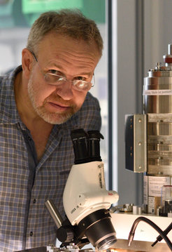
Winfried Denk and three colleagues honored with the Brain Prize
One million Euros for the invention, development and application of the two-photon microscopy
In order to fully understand structure and function of the brain, individual nerve cells and their activity must be made visible. Today, this is possible thanks to 2-photon fluorescence microscopy, which was invented and first applied to neurobiology by Winfried Denk, director at the Max Planck Institute of Neurobiology in Martinsried near Munich. The Brain Prize 2015, honoring this achievement, is awarded to Winfried Denk, Arthur Konnerth, Karel Svoboda, and David Tank. Endowed with a total of one million Euros, the award recognizes active researchers for their outstanding contribution to European neuroscience. On May 7, the prize will be presented to the scientists in Copenhagen by His Royal Highness Crown Prince Fredrik of Denmark.

Not only our mental abilities and our sensations but also our metabolism and the workings of diseases are based on processes and changes on the molecular and cellular level. These and other biological mechanisms need to be studied where they take place – within the living tissue. The necessary high-resolution observations are only possible with the aid of optical microscopes. Thus it comes as no surprise that since the early 17th century microscopes have been an integral part of scientific research. The development of fluorescent dyes has made fluorescence microscopy one of the most important technologies in today's biomedical research. With the aid of such dyes, individual cells, details of specific cells or particular cellular processes become visible under the microscope.
Multi-photon fluorescence microscopy was invented by Winfried Denk, Jim Strickler, and Watt Webb in the late 1980’s [1]. Together with his colleagues at Bell Labs (USA), including co-awardees David Tank and Karel Svoboda, Winfried Denk started to expand the use of multiphoton microscopy to neurobiological questions. Scientists from other laboratories also applied the, at that time, cutting-edge technology to brain research – co-awardee Arthur Konnerth being one of the first and most successful. Today, multi-photon microscopy, generally in the form of 2-photon fluorescence microscopy, is an essential component of most biomedical laboratories. The technique revolutionized imaging-based scientific research by enabling scientists to see deep into strongly scattering brain tissue for extended periods of time. With the aid of these microscopes, it became possible to investigate tissue layers and biological processes that were until then unreachable for imaging with an optical microscope. For the development of two-photon microscopy and its application to brain research, the four scientists are being awarded the Brain Prize 2015.
The 2-photon fluorescence microscopy
The method makes use of the interaction of two light particles (photons) with one fluorescent molecule. When two photons arrive at the same time at the location of the molecule they can excite the molecule in a "collaborative effort" by combining their quantum energies. This effect makes it possible to use less harmful long-wavelength (red or infrared) light instead of the otherwise utilized shortwave (blue or ultra-violet) light. An additional benefit of long-wavelength light is that it scatters less than shortwave light when entering the tissue. This improves the penetration depth and allows the investigation of structures that lie as deep as 1000 µm (1mm) inside the tissue instead of 50-80 µm with conventional light microscopes.
Winfried Denk
Winfried Denk was born 1957 in Munich. He studied physics at the Ludwig Maximilians University in Munich and at the ETH in Zurich (Switzerland) and obtained a PhD in Physics from Cornell University in Ithaca (USA). Then he moved to the IBM Research Lab in Rüschlikon (Schwitzerland) and later led an independent research group at Bell Laboratories in Murray Hill (USA). In 1999, Winfried Denk was appointed director at the Max Planck Institute for Medical Research in Heidelberg. Since 2011, he has been a director at the Max Planck Institute of Neurobiology, Martinsried.
Winfried Denk received numerous awards, including the Young Investigator Award of the Biophysical Society in 1998, the Rank Prize for Opto-Electronics in 2000, the Gottfried Wilhelm Leibniz Prize of the German Research Society (DFG) in 2003, the Kavli Prize in 2012, and the Rosenstiel Prize of Brandeis University in 2014. He was elected a foreign associate of the National Academy of Sciences (NAS) in 2013 and a member of the European Molecular Biology Organization (EMBO) in 2014.
More recently Winfried Denk has turned his attention from imaging the brain’s activity to mapping is circuit diagrams. To this end he developed the serial block-face electron microscope and used it to generate a precise map of all nerve cells and their connections in a piece of mouse retina [2]. This example of combining cutting-edge imaging and data analysis revealed new cell types and unknown circuit motifs (see research news "The brain: with all its cells and their connections").


