EM-Histo Lab
The service team of the EM-Histo lab assists the institute's scientists by producing histological preparations for light- and electron-microscopic analyses. We help our scientists and guests with the design, implementation and analysis of their projects. Our technical know-how and the provision of equipment and chemicals ensure a quick and efficient realization of scientific projects. If desired, we run complete projects on behalf of the scientists. In addition, we also provide training opportunities for the use of our instrument, enabling the scientists to realize their own projects.
Equipment
- Paraffin microtome: Reichert-Jung Supercut 2065
- Cryostat: Leica CM 3050
- Vibrating-blade microtome: Leica VT1000S
- Ultramicrotomes: Leica UC6, Reichert-Jung Ultracut E
- Microscopes: Zeiss Axiophot mit Diskussionstubus und digitaler Kamera Leica DFC 490, 2 Stereomikroskope (Leica M80, Wild M3)
- Stainer: Leica Ultrostain
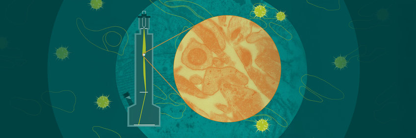
- TEM: JEOL Transmissions-Elektronenmikroskop (JEM-1230, 40-120 KV), Gatan Orius CCD Kamera
- Misc.: Leica EM Trimm
Techniques
- Histology: Paraffin sectioning, Cryostat sectioning, Vibratome sectioning, Histological staining, Immunohistochemical staining
- EM: Processing and embedding of specimens, Semithin and ultrathin sectioning, Serial sectioning, Immunolabeling, Image processing, Reconstruction