Mechanisms of Direction Selectivity

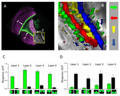
The fly visual system consists of four successive visual neuropile layers each built from repetitive columns arranged in a retinotopic way: the lamina, the medulla, the lobula and the lobula plate. Fly photoreceptors R1-6 synapse, either directly or indirectly, onto different lamina neurons, which then form the inputs to two parallel channels for the analysis of motion (Joesch et al., 2010): one circuit is specialized to detect motion of brightness increments (ON-channel), the other circuit to detect motion of brightness decrements (OFF-channel). T4 and T5 cells form the output of each channel, respectively. There are four T4 and four T5 cells per column, tuned to the four cardinal directions: front-to-back, back-to-front, upwards and downwards. According to their preferred direction, T4 and T5 cells terminate in four different layers of the lobula plate (Maisak et al., 2013). There, T4 and T5 cells make direct cholinergic, excitatory connections onto the dendrites of the tangential cells, as well as onto lobula plate interneurons, which inhibit the tangential cells in the adjacent layer (Mauss et al, 2014; Mauss et al, 2015).

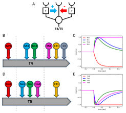
Within the motion pathway, T4 and T5 neurons are the first cells that respond to motion in a directionally selective way. Direction selectivity in T4 and T5 cells is based on three inputs: a delayed amplifier on the preferred side, a fast central exciter, and a delayed inhibitor on the null side. The amplifier enhances the central signal for motion along one direction (the ‘preferred direction’), while the inhibitor blocks the central signal during motion along the opposite direction (the ‘null direction’). Such a dual mechanism insures a high degree of direction selectivity that is observed right at the first stage where this important quantity is computed (Haag et al. 2016, 2017). In nice agreement with this result from physiology, connectomic analysis of the motion pathway reveals a tripartite dendrite with one cell on the preferred side, and several cells in the center as well as on the null side (Shinomiya et al, 2019). Interestingly, the dynamics of the different input neurons fit this scheme by exhibiting low-pass characteristics for those cells that synapse on the preferred and the null side, and band-pass characteristics for those cells that synapse onto the central part of the dendrite of T4 and T5 cells (Arenz et al, 2017). Current projects concern the biophysical mechanism by which one signal amplifies another signal within the dendrite as well as the question why T4 and T5 cells receive input from so many different cells, and not just three.
References
Groschner lN, Malis JG, Zuidinga B, Borst A (2022) A biophysical account of multiplication by a single neuron. Nature 603: 119-123.
Arenz A, Drews MS, Richter FG, Ammer G, Borst A (2017) The temporal tuning of the Drosophila motion detectors is determined by the dynamics of their input elements. Curr Biol 27: 929-944.
Haag J, Arenz A, Serbe E, Gabbiani F, Borst A (2016) Complementary mechanisms create direction selectivity in the fly. eLife 5: e17421.
Haag J, Mishra A, Borst A (2017) A common directional tuning mechanism of Drosophila motion-sensing neurons in the ON and in the OFF pathway. eLife 6: e29044.
Joesch, M., Schnell, B., Raghu, S. V., Reiff, D. F. & Borst, A. ON and OFF pathways in Drosophila motion vision. Nature 468, 300–304 (2010).
Maisak, M. S. et al. A directional tuning map of Drosophila elementary motion detectors. Nature 500, 212–216 (2013).
Mauss AS, Meier M, Serbe E, Borst A (2014) Optogenetic and pharmacologic dissection of feedforward inhibition in Drosophila motion vision. J Neurosci 34: 2254-2263.
Mauss AS, Pankova K, Arenz A, Nern A, Rubin GM, Borst A (2015) Neural circuit to integrate opposing motions in the visual field. Cell 162: 351-362.
Shinomiya K et al (2019) Comparisons between the ON- and OFF-edge motion pathways in the Drosophila brain. eLife 8: e40025.

